
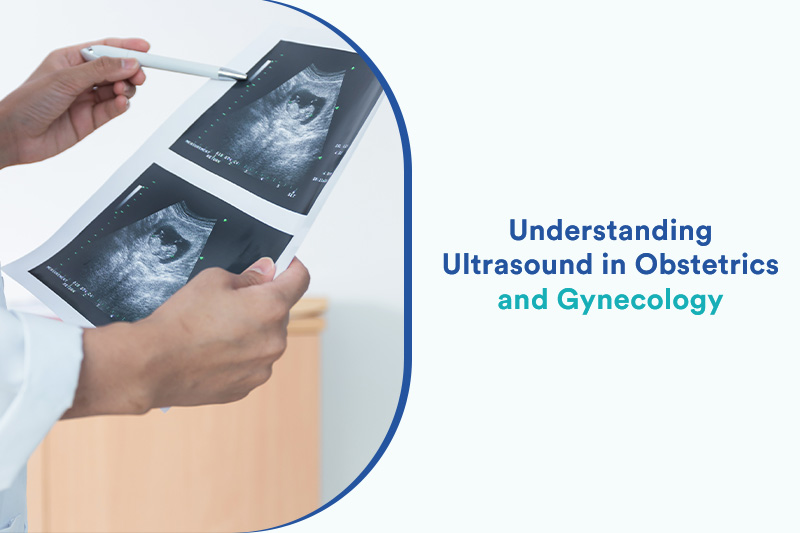
Ultrasound is an imaging technique that uses sound waves to create images of the internal organs and other structures in your body. Introduction of ultrasound has significantly impacted patient care as due to this imaging of fetus and placenta in obstetrics and maternal organs in gynecology is possible with high resolution and it has ultimately benefitted in advanced diagnosis and treatment. A number of illnesses and ailments may be diagnosed, and therapy can be planned by these images. Ultrasound frequencies commonly used in the OBGYN are between 3 to 10 MHz. Without intrusive procedures, ultrasound can assist in precisely examining the interior organs. It is a medical examination carried out by a physician to examine your body’s interior organs in order to assess or identify the disorder’s underlying cause. B-mode display, which stands for “Brightness mode” i.e., 2D imaging, is mainly used to describe any form of grey scale display of ultrasound image.
The routine obstetric ultrasound scan offers a precise and secure clinical evaluation of the gravid uterus during a woman’s pregnancy, including detecting the number of embryos present, characterising the pregnancy location, and assisting in the prenatal diagnosis of foetal defects. Thus, Ultrasound uses sound frequencies and works as a screening and diagnostic tool in the medical field.
It is highly important for every medical professional to know the basic obstetrics ultrasound examination and ultrasound scanning techniques, such as:
- Selecting the appropriate transducer and settings
- Apply minimal pressure on the abdomen
- Reduce depth to a minimum
- Minimize the sector width
- Adjust focal zones
- Zoom area of interest
- Maintain the target anatomic area in the centre of screen
Obstetrics and Gynecology ultrasound is often employed in the following:
- evaluating the pelvic organs
- identifying acute appendicitis
- in diagnosis and treatment of gynecologic issues such as, endometriosis, leiomyoma, adenomyosis, ovarian cysts, and lesions
- recognising adnexal masses, such as an ectopic pregnancy,
- diagnosing gynecologic cancer
- monitoring the ovarian follicles’ reaction to fertility drugs like Pergonal during infertility therapy. But it frequently underestimates the actual ovarian volume
- Diagnosis and management of twin pregnancies
- Diagnosis of co-joined twins can be made in the made trimester by grey scale and colour Doppler ultrasound
- Dating and growth monitoring
The objectives of Ultrasound Examination includes:
- Confirmation of pregnancy
- Single vs multiple pregnancy
- Intrauterine localisation of gestational sac
- Assessment of basic anatomy
- Monitoring the progress of fetal development
- Knowing reproductive disorders
To enhance your skills and excel in the field of OBGYN, enrolling in one of the best online certification courses on Ultrasound in OBGYN is a good option. A good online ultrasound course will provide you with in-depth knowledge on the clinical applications of the academic knowledge that will help hone gynecology skills. ‘Ultrasound in OBGYN made easy’ is one of the best online courses available on ultrasound. This course is the finest approach to educate students the fundamentals of ultrasonography while laying the groundwork for opening an independent clinic thanks to its outstanding lectures, thorough notes, and pioneering teachers. This training is designed to give medical professionals advice on how to identify foetal anomalies in ultrasounds and other patients with gynaecological issues.
An aspirant must learn the correct way of making the right choice regarding scanner, probes and softwares, optimizing the 2D Ultrasound images, basics of fetal echocardiography, systematic transvaginal scan, role of ultrasound in infertility practice, doppler in PIH and FGR, and other important topics and gain practical knowledge.


